The Biology Corner: Sheep Brain Dissection
| Record number: | 15639 |
|---|---|
| Category: | Dissection |
| Type: | Web pages |

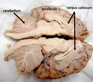
Sheep Brain Dissection shows images of the structures that are visible during a sheep dissection, where some of the photos are labeled. Includes structures like the lobes of the cerebrum, the cerebellum with arbor vitae, the pituitary and the brain stem (medulla, pons, and spinal cord). Internal structures include the corpus callosum, lateral ventricle, fornix, superior and inferior colliculi, pineal gland, thalamus and hypothalamus.
Students can use the Sheep Brain Dissection Guide, Label the Brain of the Sheep & Cranial Nerves Coloring through the dissection procedure.
Images of other dissection procedures from The Biology Corner: Sheep Heart Dissection, Squid Dissection, Fetal Pig Dissection, Frog Dissection & Rat Dissection.
For comparison please see:
- Sheep Brain Dissection: The Anatomy of Memory
- The Sheep Brain Dissection Guide
- The Sheep Brain: A Basic Guide
- The Concise Brain/ Heart
Access online.
Takk for din tilbakemelding! Vær oppmerksom på at vi ikke kan kontakte deg hvis ikke du oppgir din epostadresse.
Hva lette du etter?
Gi oss gjerne en tilbakemelding slik at vi kan forbedre informasjonen på siden. På forhånd takk for hjelpen! Vennligst skriv inn din epostadresse hvis du vil ha et svar.
Kontakt oss gjerne på e-post hvis du har spørsmål.
