The Biology Corner: Sheep Heart Dissection
| Record number: | 6daa8 |
|---|---|
| Category: | Dissection - Education and training |
| Type: | Web pages |

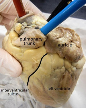
Sheep Heart Dissection shows images of all of the structures that are visible during a sheep heart dissection, such as aorta, pulmonary artery, pulmonary vein, and the vena cava.
Students can use Sheep Heart Dissection through the dissection procedure. The guide includes instructions on how to determine which side of the heart is the ventral (front) side by searching for the auricles and the interventricular sulcus. Students are instructed to cut the heart lengthwise through the atrium and ventricle so that each side can be opened and compared.
More Pig dissection images can be found at: https://photos.google.com/share/AF1QipPydEBsd6LA8bDfjCdXVuQSThnbq8NOdqapFEEQ_OWcypnzDgjoyvWtNhgbVbVlyQ?key=eENIUWx3OTdSWXQ4Xy0zWVluQzBYZWwyQWs0amJ3.
Please also see Video: The Dissection of the Sheep Heart.
Images of other dissection procedures from The Biology Corner: Sheep Brain Dissection, Squid Dissection, Fetal Pig Dissection, Cow Eye Dissection, Frog Dissection & Rat Dissection.
For comparison please see:
Access online.
Takk for din tilbakemelding! Vær oppmerksom på at vi ikke kan kontakte deg hvis ikke du oppgir din epostadresse.
Hva lette du etter?
Gi oss gjerne en tilbakemelding slik at vi kan forbedre informasjonen på siden. På forhånd takk for hjelpen! Vennligst skriv inn din epostadresse hvis du vil ha et svar.
Kontakt oss gjerne på e-post hvis du har spørsmål.
