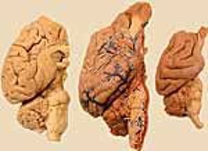
Brain Gross Anatomy
By T. F. Fletcher
| Record number: | bbaf9 (legacy id: 5352) |
|---|---|
| Category: | Anatomy |
| Type: | Web pages |

Description: Brain Gross Anatomy is a web site that presents domestic mammalian brain neuroanatomy from a gross anatomical perspective. For each brain region, neural components are listed in hierarchical order, and there are links to images of brain surfaces, brain dissections and sections of brains. All navigation links display a colour change (to red) when the mouse is moved over them. The following options are available: Navigate to major Sections of the web site, including the Home page, by clicking an available Section option listed at the top of the page, just below the banner; Within a major Section, navigate to a particular brain region/component by clicking a pertinent link term (link terms are blue text terms terminated by ellipses). Image display: Clicking a display option (blue text terminated by a colon) will display a pop-up window containing a pertinent image & caption; additional pop-up images may be displayed by clicking the small images arranged along the left side of a page; Within a pop-up image window, click "LABELS" to toggle labels on/off or "Return" to close the window (or use the operating system to close windows). Includes: Brain Divisions; Cranial Nerves; Ventricles & Vessels. Images of different animal brains with labels are presented (dog, cat, sheep and horse). The Brain Divisions explains 3 different ways to subdivide the brain: 1 Embryonic divisions: Telencephalon (Cerebrum); Diencephalon; Mesencephalon; Metencephalon; Myelencephalon; 2 Gross Anatomy divisions: Cerebrum (telencephalon); Cerebellum (part of the metencephalon); and Brainstem (di-, mes-, pons, myel-encephalon); 3 Clinically useful divisions: Forebrain (telencephalon & diencephalon) - mental status: voluntary movements and vision; and Hindbrain (metencephalon & myelencephalon) - cerebellum & ventricular syndromes and nerve deficits. In the Cranial Nerves section, all 12 cranial nerves are described. In the Ventricles section all 4 ventricles and the central canal with spinal cord are described, and in the Vessel section the internal carotid arteries are described.
Comments & References:
Suitable for first-year veterinary students studying CVM 6120 Veterinary Neurobiology. The Veterinary Anatomy website at the University of Minnesota has been redesigned. Please check the homepage for more information on the availability of this product.
Computer type: Macintosh, IBM
Price: Free of charge
Free of charge Free of charge
Takk for din tilbakemelding! Vær oppmerksom på at vi ikke kan kontakte deg hvis ikke du oppgir din epostadresse.
Hva lette du etter?
Gi oss gjerne en tilbakemelding slik at vi kan forbedre informasjonen på siden. På forhånd takk for hjelpen! Vennligst skriv inn din epostadresse hvis du vil ha et svar.
Kontakt oss gjerne på e-post hvis du har spørsmål.
