Lab1: Nervous Tissue Histology
By Thomas F. Fletcher
| Record number: | 01170 (legacy id: 5702) |
|---|---|
| Category: | Histology |
| Type: | Web pages |

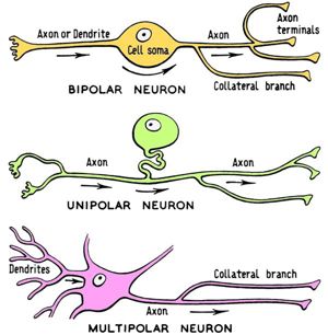
This web site reproduces Lab1: Nervous Tissue Histology from the Lab Manual for CVM 6120 Veterinary Neurobiology. The site was established to display lab images in colour with descriptive text. Neurohistology Lab Objectives: A. Identify neurons, astrocytes, oligodendrocytes, and ependymal cells in the Central Nervous System (CNS); B. Identify axons, myelin, and lemmocytes associated with nerves and ganglia in the PNS; C. Be able to distinquish white matter (collections of myelinated axons) from grey matter (accumulations of neuronal cell bodies) in the CNS; D. Be able to identify dura mater, arachnoid, subarachnoid space, and pia mater; E. Be able to define the following terms: 1 multipolar neuron; 2 bipolar neuron; 3 unipolar neuron; 4 perikaryon; 5 Nissl substance; 6 axon; 7 dendrite; 8 telodendria. Neurohistology Topics: Neurons; Neuroglial cells; Peripheral nerves; Spinal cord and meninges; Spinal ganglia; Receptors (sensory endings) - lamellar (Pacinian) corpuscle. Please also view the Neurohistology Atlas Web Site.
Comments & References: The Veterinary Anatomy website at the University of Minnesota has been redesigned. Please check the homepage for more information on the availability of this product. Also see record number 5355 for information on other Neurobiology Labs.
Price: Web pages: Free of charge
Version: 2004
Free of charge Web pages: Free of charge
Thanks for your feedback! Please note that we cannot reply to you unless you send us an email.
What are you looking for?
We value your feedback so we can improve the information on the page. Please add your email address if you would like a reply. Thank you in advance for your help.!
Please contact us by email if you have any questions.
