Atlas on Veterinary NeuroHistology
By Thomas F. Fletcher
| Record number: | 22f6b (legacy id: 5705) |
|---|---|
| Category: | Histology |
| Type: | Web pages |

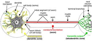
This web site presents an atlas of nervous tissue images with captions, along with an image catalogue. This atlas is divided into two sections: Neurohistology Atlas Images; and Neurohistology Atlas Catalogue. In the Atlas Images section, the viewer may choose between: Neurons & synapses (1 image); Neuroglia & myelin (1 image); CNS: white & grey matter (1 image); PNS: nerves, ganglia & receptors (1 image); Meninges (1 image). In the Atlas Catalogue section, the viewer may choose between: Neurons & synapses (14 images); Neuroglia & myelin (17 images); CNS: white & grey matter (11 images); PNS: nerves, ganglia & receptors (16 images); Meninges (5 images). Each image in both sections is followed by a descriptive text.
Comments & References: Suitable for student self-study. This web site may be viewed online or downloaded to run from a local hard disc. It is recommended that first-time viewers should choose "Instructions and Download Info".
The Veterinary Anatomy website at the University of Minnesota has been redesigned. Please check the homepage for more information on the availability of this product.
Price: Free of charge
Version: 2004
Free of charge Free of charge
Thanks for your feedback! Please note that we cannot reply to you unless you send us an email.
What are you looking for?
We value your feedback so we can improve the information on the page. Please add your email address if you would like a reply. Thank you in advance for your help.!
Please contact us by email if you have any questions.
