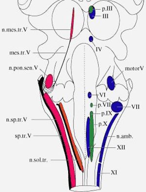
Cranial Nerve Nuclei 1.2
By T. F. Fletcher
| Record number: | 0c5bb (legacy id: 5348) |
|---|---|
| Category: | Anatomy - Histology |
| Type: | Computer Program |

Description: Cranial Nerve Nuclei 1.2 is an independent study courseware intended to help veterinary students learn neuroanatomy of cranial nerve nuclei in the dog brain. Cartoons of cranial nerve cell columns, colour-coded by type (SE, VE, GSA, GVA, etc.), are shown in dorsal, transverse, and sagittal views of a canine brainstem. Cranial nerve nuclei are also labelled in transverse sections through a canine brain. This courseware has five sections: 1 Introduction to general features of afferent & efferent and somatic & visceral nuclei; 2 Illustrations of cranial nerves plus a synopsis of innervation & fiber-type per cranial nerve; 3 Neurological deficits resulting from damage per cranial nerve; 4 General afferent and efferent cell columns and the cranial nerve nuclei they form; 5 Special afferent pathways and nuclei (vision, hearing, kinesthesia, taste and olfaction).
Comments & References: Suitable for first year veterinary students studying neurobiology. Cranial Nerve Nuclei 1.2 is a SuperCard stand-alone application (Mac platform). Minimum hardware requirements: Macintosh computer with a colour display, optimised for 640 x 480 pixel resolution and thousands of colours. Requires 10 MB RAM. Disc size: 35 MB. The Veterinary Anatomy website at the University of Minnesota has been redesigned. Please check the homepage for more information on the availability of this product.
Computer type: Macintosh
Price: Free of charge
Version: 1.2, 1999
Free of charge Free of charge
Thanks for your feedback! Please note that we cannot reply to you unless you send us an email.
What are you looking for?
We value your feedback so we can improve the information on the page. Please add your email address if you would like a reply. Thank you in advance for your help.!
Please contact us by email if you have any questions.
